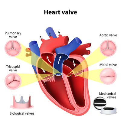
 |
| Image © Designua | Dreamstime.com |
A Tribute
A decade has passed
since my mitral valve repair.
Who would have surmised
I would still be here! Kudos
to Doctor Gammie and Team!
Verse is Tanka: 5,7,5,7,7 Syllabic content
Click on "Heart: Mitral Valve Repair" post in this Blog to check out details of Humble Blogger's Mitral valve repair at the University of Maryland Medical Center in Baltimore on 5/11/2006. A momentous time in my life!
Currently my heart is in atrial fibrillation (A-Fib) about 80% of the time even though just before my mitral valve repair, heart surgeon Dr. Gammie performed a cryogenic (cold) MAZE (ablation) procedure for amelioration of the atrial fibrillation. The procedure wasn't 100% effective but my A-Fib is now controlled with the beta blocker Metoprolol and (of course) warfarin to increase blood clot time. I've toyed with the idea of fine tuning the ablation treatment but so far have done nothing additional. Unfortunately I was not taking Warfarin when I suffered a stroke on 9/12/2012, from which I've recovered quite well :) See link in this blog "Time to Say Goodbye"
Below are results from a cardiac ECHO done 11/3/2015:
Transthoracic Echocardiography Report (TTE)
Patient Name KUBIATOWICZ DAVID Date of Study 11/03/2015
Date of Birth 04/18/1942 Referring Physician ARDOLF, JOSEPH C
Age 73 year(s)
Gender Male Interpreting HEHC OUTPATIENT
Physician JOHNSON, THOMAS MD
Procedure
TTE procedure: ECHO COMPLETE.
Procedure Date
Date: 11/03/2015 Start: 12:37 PM
Study Location: St Johns
Technical Quality: Adequate visualization
Patient Status: Routine
Height: 69 inches Weight: 189 pounds BSA: 2.02 m^2 BMI: 27.91 kg/m^2
HR: 67 bpm BP: 125/85 mmHg
Indications
Mitral valve disorder, atrial fibrillation and aortic aneurysm.
Conclusions
Summary
Normal left ventricular size.
Normal left ventricle wall thickness.
Left ventricular ejection fraction is visually estimated to be 60%.
No regional wall motion abnormalities.
Normal right ventricular size and systolic function.
Mild dilation of ascending aorta .
An annuloplasty repair is noted in the mitral position.
Posterior leaflet with marked reduced excursion c/w repair.
Mild mitral regurgitation is present.
No evidence of mitral valve stenosis.
Findings
Left Ventricle
Normal left ventricular size.
Normal left ventricle wall thickness.
Left ventricular ejection fraction is visually estimated to be 60%.
No regional wall motion abnormalities.
Right Ventricle
Normal right ventricular size and systolic function.
Left Atrium
Moderately enlarged left atrium.
Right Atrium
Moderately enlarged right atrium.
Great Vessels
Mild dilation of ascending aorta .
Aortic Valve
Tricuspid aortic valve.
No evidence of aortic stenosis.
Mitral Valve
An annuloplasty repair is noted in the mitral position.
Posterior leaflet with marked reduced excursion c/w repair.
Mild mitral regurgitation is present.
No evidence of mitral valve stenosis.
Tricuspid Valve
Mild tricuspid regurgitation.
Estimated right ventricular systolic pressure is 32 mmHg.
Pulmonic Valve
The pulmonic valve is not well visualized.
Mild pulmonic regurgitation is present.
No evidence of pulmonic valve stenosis.
Pericardial Effusion
No pericardial effusion.
Signature
----------------------------------------------------------------
Electronically signed by JOHNSON, THOMAS MD(Interpreting
physician) on 11/03/2015 03:27 PM